
A painful or swollen leg and a breathing emergency may seem unrelated. They can be connected by the circulation: a clot that forms in a deep vein can shed a fragment that reaches the lungs. The resulting blockage is called a pulmonary embolism, or PE. It is a potentially life-threatening condition that needs immediate medical attention. Source: NHLBI pulmonary embolism overview.
Sudden difficulty breathing, chest pain, or fainting needs emergency assessment. Contact your local emergency service; do not wait for leg swelling or try to diagnose the cause from this article. These symptoms can also occur in other serious conditions. Source: NHS emergency guidance.
Watch the 42-second explanation
Where the problem can begin: a deep vein
Deep vein thrombosis, or DVT, means that a clot has formed in a deep vein. The leg is a common location, although clots can also develop elsewhere. In the affected limb, possible signs include swelling, pain or tenderness, warmth, and a change in skin colour. Some DVTs cause no noticeable symptoms. New symptoms that suggest DVT should be assessed promptly rather than watched indefinitely. Source: CDC blood-clot overview.
A DVT and a PE describe different parts of the problem. DVT names a clot in a deep vein. PE names a blockage in the lung circulation. One can lead to the other, but a clot in a leg does not inevitably travel to the lungs. The film uses one illustrative journey to explain what can happen; it does not predict what will happen to an individual.
The route: leg vein, right heart, pulmonary artery
Veins return blood towards the heart. If a piece of a leg-vein clot detaches, the blood can carry it through larger veins, into the right atrium and right ventricle, and then into the pulmonary arteries. These arteries carry blood from the right side of the heart towards the lungs. The fragment may lodge in a vessel where it obstructs blood flow. Source: American Thoracic Society patient information.
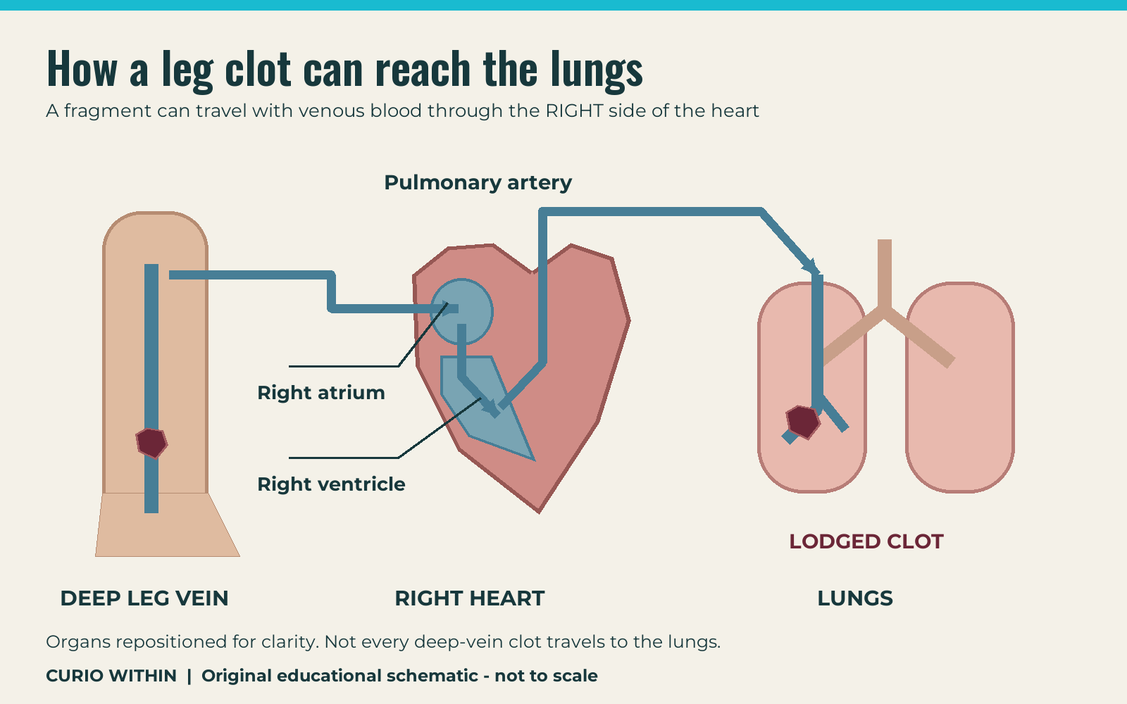
The right-heart step is essential to understanding the journey. This is a venous clot entering the lung circulation, rather than a diagram of a clot travelling through the left heart to the brain. The severity of a PE depends partly on how much lung blood flow is obstructed. A large obstruction can also put extra strain on the right side of the heart. Source: ATS explanation of circulation and heart strain.
Why breathing can become difficult even if air gets in
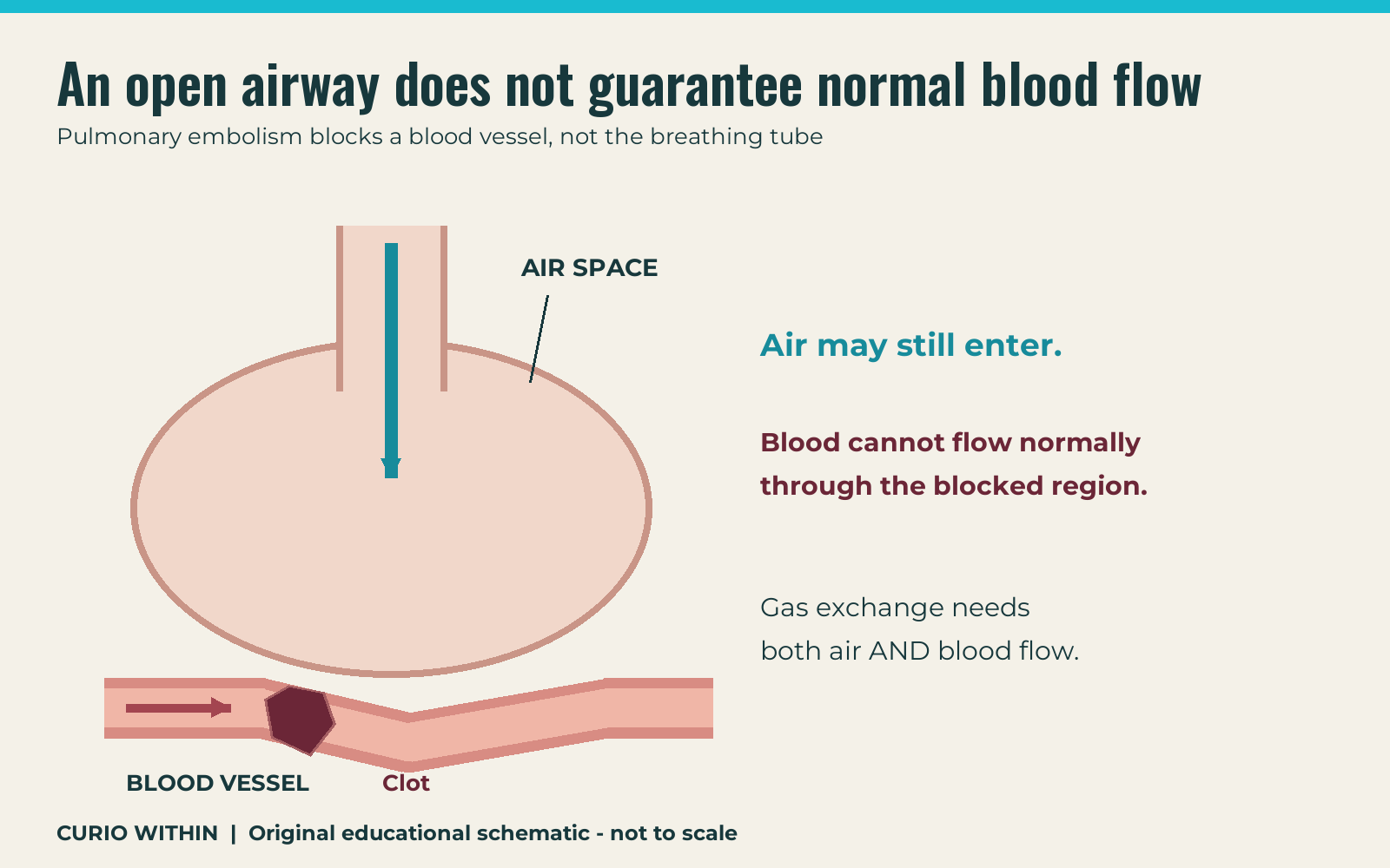
Breathing brings air into the lungs, but oxygen also needs blood to carry it away. A pulmonary embolism blocks an artery supplying part of the lung. Air may still reach the affected air spaces while blood flow is reduced or stopped in their neighbouring vessels. That mismatch can interfere with gas exchange. The obstruction is in a blood vessel, not a piece of clot plugging the windpipe. Source: NHLBI explanation of pulmonary arterial blockage.
Warning signs are not limited to the leg
PE can cause sudden breathlessness, pain that gets worse when taking a deep breath, or coughing up blood. A person may also feel lightheaded or faint. Importantly, PE can occur without obvious DVT symptoms first. A normal-looking leg is therefore not a reliable way to rule it out. Source: CDC signs and symptoms.
Do not wait to see whether all the signs appear together. Sudden breathing difficulty or chest pain warrants emergency help, even when there is no swollen calf. The NHS also advises against driving yourself to emergency care when PE is suspected. Follow the instructions of your local emergency service. Source: NHS advice on getting help.
How clinicians investigate a suspected clot
Symptoms alone cannot confirm PE. A clinician considers the medical history, examination, risk factors and the most appropriate tests. Ultrasound is commonly used to look for DVT in a limb. Investigations for possible PE can include a CT pulmonary angiogram or, in selected circumstances, a ventilation-perfusion scan. Blood tests may help guide assessment, but their results must be interpreted in context. A D-dimer result is not a stand-alone diagnosis. Source: NHLBI diagnosis information.
Treatment is a medical decision, not a home experiment
Anticoagulants are often called blood thinners. They help stop clots growing and reduce the formation of new clots; they do not simply melt an existing clot instantly. Some severe cases require other medicines or procedures. The appropriate approach depends on the clinical situation, including bleeding risk. Do not start, stop or change a clotting-related medicine because of a short video or an online diagram. Source: NHLBI treatment overview.
The idea to remember
The connection is a route through the circulation: deep leg vein, right heart, pulmonary artery. An obstruction there affects blood reaching the lung, even if air can still get in. The practical takeaway is to seek emergency help for sudden breathing difficulty or chest pain, without waiting for a leg symptom to appear.
Curio Within educational explainer. Sources checked 28 October 2026. This article is general information, not an individual diagnosis or treatment plan. Illustrations are simplified original schematics. No clinical review is claimed.
Keep your curiosity going
Why Asthma Makes Breathing Hard: Inside a Narrowing Airway
See how tight airway muscles, swelling and extra mucus can make breathing harder during asthma, and learn when symptoms need emergency care.
Read the story →
What Actually Happens Inside Your Brain During a Stroke?
One blocked artery can disrupt a brain network. Follow the chain from blood flow to neurons, then learn why possible stroke signs need emergency care.
Read the story →