
A stroke can suddenly affect speaking, movement or vision. The cause begins inside the brain’s blood supply, and its consequences reach individual cells. In an ischemic stroke, an artery becomes blocked and the tissue it supplies receives too little blood. This article follows that type of stroke from an artery to a neuron, then explains why urgent medical assessment matters. NHLBI: What is a stroke?
Possible stroke symptoms are an emergency. Call your local emergency number immediately, including when symptoms improve.
Watch the visual explanation
Blockage and bleeding are different problems
The two main stroke categories are ischemic and hemorrhagic. Ischemic strokes involve inadequate blood flow, commonly because a clot blocks an artery. Hemorrhagic strokes involve bleeding from a vessel into or around brain tissue. Both can injure the brain, but treatment is different. The video follows one artery-blockage example rather than presenting every stroke as the same event. NINDS: Stroke overview
An embolus is a clot or other obstructing material that travels from elsewhere and lodges in a vessel. A thrombus can instead form locally. The animation uses a travelling blood clot. It is a soft aggregate of blood components, not a stone bouncing through an artery.
Follow the supply route
The middle cerebral artery, or MCA, supplies much of the brain’s lateral surface and important deeper structures through its branches. Its territory includes regions involved in movement, sensation and language. We use a left-sided MCA example, while recognizing that branching patterns and the affected territory vary. NCBI: Middle cerebral artery anatomy
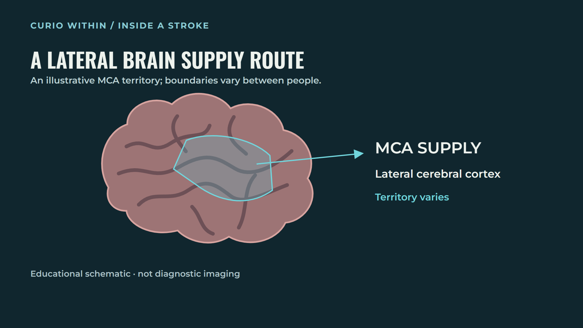
This is an explanatory map, not a patient’s scan or a precise prediction of injury.
Arteries divide into smaller vessels and eventually a dense capillary network. Blood carries oxygen and glucose close to neurons and supporting cells. The cells use these supplies to support energy production. Blood normally remains within the vessels; it does not wash directly over neurons. A microscopic cutaway enlarges the relationship so it can be seen. NIH: Stroke and related effects
What changes inside a threatened neuron?
Neurons maintain different concentrations of ions across their membranes. Those differences help make electrical signaling possible. Maintaining them requires energy-dependent transport, including the sodium–potassium pump. Severe interruption of oxygen and glucose supply undermines energy production. Pump function can no longer keep pace, ions redistribute and water movement contributes to cell swelling. Ionic regulation and cell-volume changes after stroke; Disrupted ionic homeostasis
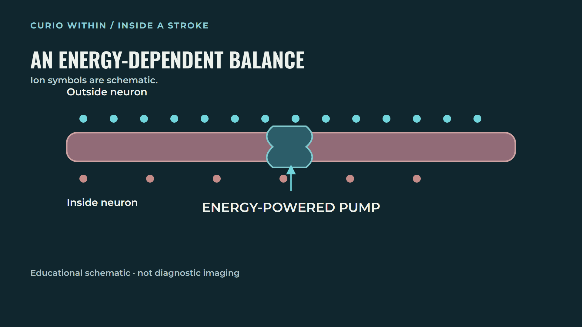
The membrane and ion symbols are enlarged. Their size and number are not measurements of a real cell.
Mitochondria are part of the cell’s energy machinery. Ischemia can disturb both their function and structure. Loss of usable energy, membrane instability and mitochondrial injury interact, making the cell increasingly vulnerable. This is why an artery problem becomes more than a simple plumbing blockage. Mitochondria in ischemic stroke
When communication contributes to injury
Glutamate normally helps transmit signals across synapses. Under severe ischemic conditions, excessive glutamate can accumulate outside cells. Overstimulation of receptors and disturbed calcium regulation can activate damaging cellular processes. Membranes, proteins and mitochondria can be affected. These processes overlap; the film’s sequence is a teaching device rather than a universal order in which every neuron dies. Basic Neurochemistry: Hypoxia-ischemia; Salaudeen and colleagues, 2024
The dots used for neurotransmitters and ions are symbols. Real neurons do not produce the large glowing sparks sometimes used in entertainment animations, and blood cells do not enter the synaptic cleft.
Core and penumbra: why the damage is uneven
Blood flow does not fall to the same level throughout an affected region. Tissue exposed to very severe ischemia can become irreversibly injured. Other tissue may retain some supply, including through alternative vascular routes. It may stop functioning normally while still being potentially salvageable. This threatened tissue is often described as the ischemic penumbra. NCBI: Stroke imaging
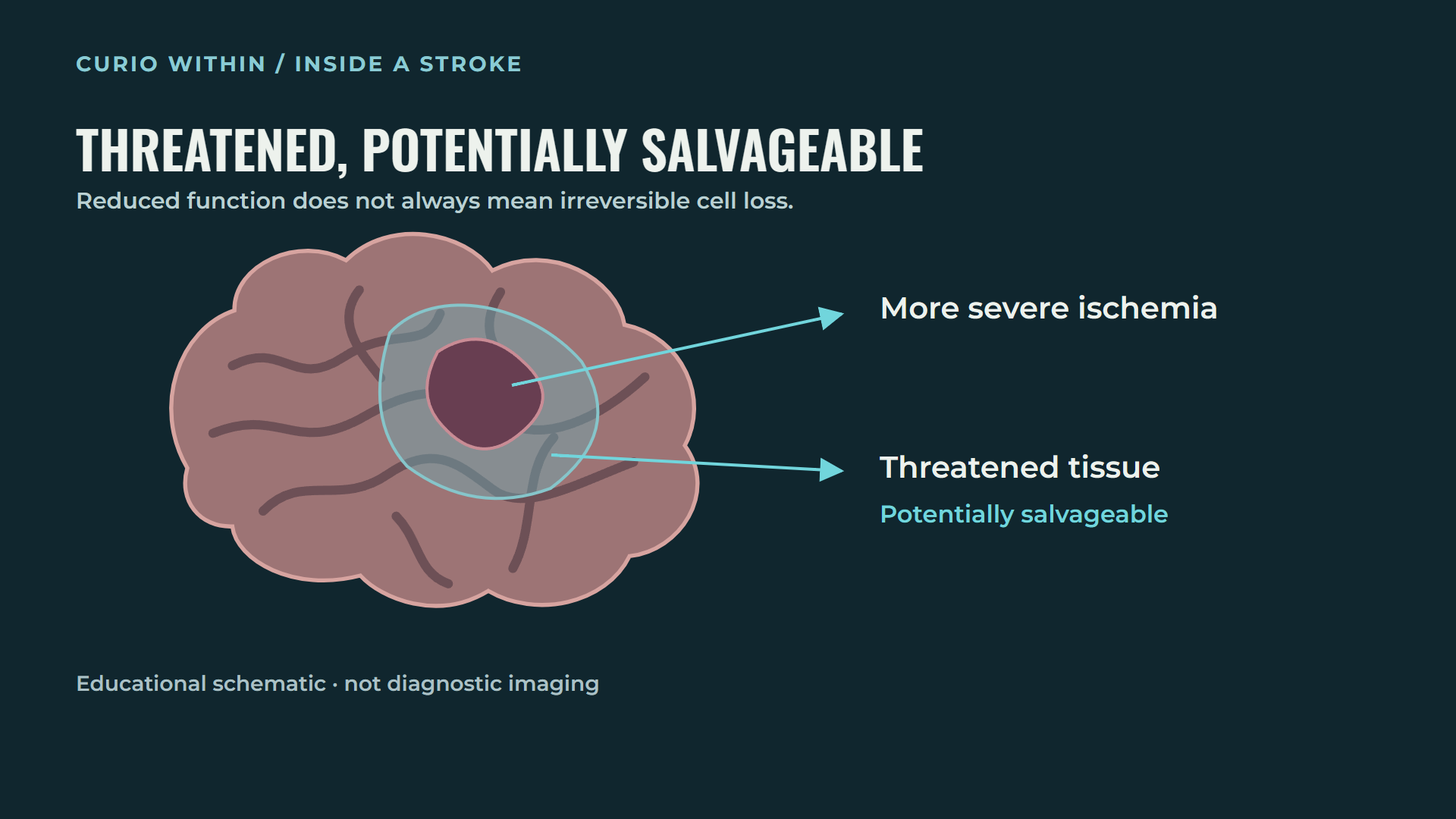
The zones are schematic. Their boundaries, size and survival vary; they are not fixed rings or a countdown.
This distinction explains the aim of rapid assessment and appropriate treatment: protect tissue that is still threatened. It does not mean that any particular person has a guaranteed amount of time remaining. The vessel involved, remaining flow, collateral routes and individual biology all matter.
Why symptoms can appear so suddenly
The brain performs tasks through connected networks. A stroke affecting motor pathways can cause weakness, often on the opposite side of the body. Damage to language networks in the dominant hemisphere—commonly the left—can interfere with speaking or understanding. Other locations can affect balance, sensation or vision. A small area of injury can therefore disrupt a task that depends on many connected regions. NINDS: Stroke overview
How clinicians decide what to do
Examination and brain imaging help distinguish ischemic stroke from bleeding and identify the affected area. CT or MRI may be used, sometimes with additional vessel or perfusion imaging. A normal-looking early scan does not by itself settle every question; the medical team combines the scan with symptoms, examination and timing. NHLBI: Stroke diagnosis; NINDS: Assess and treat
Some patients qualify for medicine that helps dissolve a clot. Selected large-vessel blockages may be treated by thrombectomy, in which a catheter-based device retrieves or aspirates the clot. The animation shows a temporary retrieval device being removed with the clot. It should not be confused with leaving a permanent coronary stent in place. Eligibility depends on the clinical findings and risks. NHLBI: Stroke treatment
Recovery and the action that matters now
Restoring blood flow does not instantly replace dead neurons. Rehabilitation can help people regain skills and support adaptation in surviving networks. Some people recover extensively; others have lasting difficulties. Recovery varies with the injury and the individual. NHLBI: Stroke recovery; NINDS: Recovery research
Sudden face drooping, arm weakness or speech difficulty are important warning signs. Sudden balance or vision changes also matter. Call your local emergency number, note when the person was last known well, and do not wait for pain or for every sign to appear. Improving symptoms still require urgent assessment. American Stroke Association: Warning signs
The central idea is that a blocked supply route can disrupt the function of individual cells and entire networks. Fast medical care aims to preserve threatened brain tissue before more injury becomes irreversible.
Educational information and simplified medical visualization, not an individual diagnosis or treatment plan. Sources checked 28 October 2026.
Keep your curiosity going
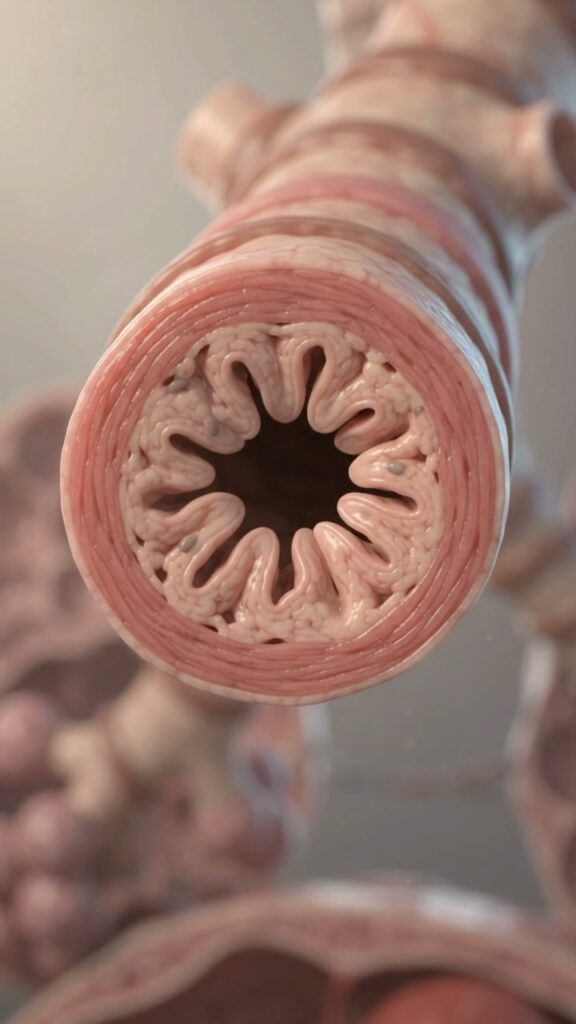
Why Asthma Makes Breathing Hard: Inside a Narrowing Airway
See how tight airway muscles, swelling and extra mucus can make breathing harder during asthma, and learn when symptoms need emergency care.
Read the story →
What Actually Happens Inside Your Heart During a Heart Attack?
The heart can keep beating while a blocked coronary artery deprives part of its muscle of oxygen. Follow the chain from artery to cell.
Read the story →