
A heart attack is often imagined as a heart suddenly switching off. Usually, the heart keeps beating. The immediate problem is that an area of heart muscle loses enough of its own blood supply to become injured. This article follows one common example: a plaque breaks inside a coronary artery, a clot grows over it, and the muscle downstream receives too little oxygen. Other kinds of heart attack also occur. American Heart Association; NHLBI.
Watch the full visual explanation
First, locate the coronary arteries
The heart pumps blood to the rest of the body, but its muscle also needs blood. Coronary arteries run along its surface and give smaller branches to the working muscle. In the film, we use the left anterior descending artery, or LAD, as a specific visual example. It descends from near the heart’s base along a groove on the front toward the apex. Its branches supply much of the front wall of the left ventricle and part of the dividing wall called the septum. The exact area affected by a blockage depends on the artery and the site of obstruction. Cleveland Clinic, LAD anatomy; NCBI Bookshelf, LAD anatomy.
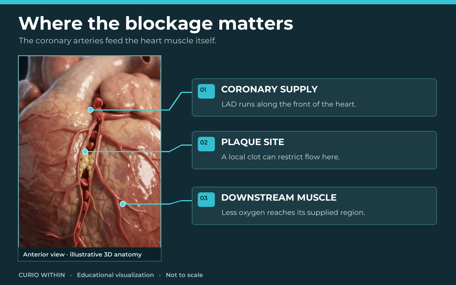
Illustrative 3D anatomy with educational callouts. This is not a map of any individual’s heart attack.
The trigger: a break in plaque
An atherosclerotic plaque develops within an artery wall over time. It contains fats, cholesterol and other material under a covering. A vulnerable area can break open. Platelets and other components of the blood collect at the exposed surface and form a clot. This clot grows from the damaged site; it is not a loose stone that suddenly appears in the middle of the artery. Depending on its size, it can partly or completely block the channel through which blood normally flows. American Heart Association; NHLBI causes.
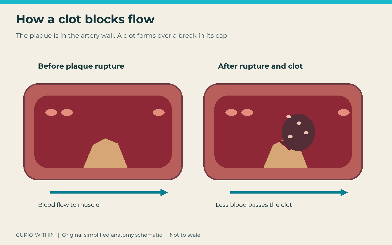
The plaque is in the wall. The clot forms at the break. This cutaway shows relationships, not the speed or exact appearance of clot formation.
What happens beyond the blockage?
The section of heart muscle fed by that artery receives less oxygen-rich blood. Muscle cells use oxygen to make energy required for contraction and essential cell functions. With severe lack of flow, energy production falls and the affected area can contract less strongly. The rest of the heart may continue to beat. A microscopic animation can make this hard-to-see chain understandable, but its particles, timing and colours are illustrative; there are no literal glowing “energy beads” inside the cells. NIH PubMed review of acute myocardial ischemia; American Heart Association.
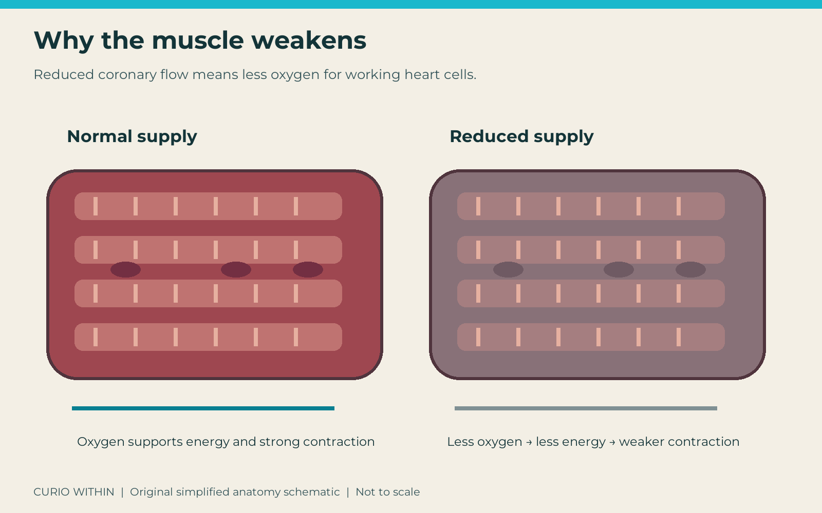
The cell rows are enlarged and simplified. A video cannot diagnose how much muscle a real patient has lost.
If blood flow remains very low or absent, more cells become irreversibly damaged. The amount of damage depends on the area supplied, the degree of blockage, other available blood routes and how quickly treatment restores flow. The film compresses a changing medical process into seconds. It does not mean tissue dies instantly when a clot forms. Damaged muscle may later heal with scar tissue; recovery varies with the person and treatment. American Heart Association; NHLBI treatment.
Heart attack or cardiac arrest?
These are different emergencies. A heart attack concerns inadequate blood supply to the heart muscle. The heart will often continue beating during it. In cardiac arrest, the heart suddenly fails to pump effectively, often because its electrical rhythm becomes dangerously disordered. A heart attack can trigger such a rhythm, but many heart attacks do not lead to cardiac arrest immediately. The distinction helps explain why a person can be awake and talking while heart muscle is being injured. Both require urgent medical care. American Heart Association comparison.
How clinicians identify the problem
The cause cannot be confirmed by watching an animation or matching a symptom list. Medical teams assess symptoms and examine the person, usually using an electrocardiogram, or ECG, to record electrical activity. Blood tests may measure cardiac troponin, a protein that can enter the blood when heart muscle is damaged. Tests may be repeated to assess changes. The findings guide treatment; not every person has the same test pattern or needs the same procedure. NHLBI diagnosis.
Why speed matters
Medical teams may use medicines or a procedure to improve blood flow. One procedure, percutaneous coronary intervention, uses a catheter to open a blocked coronary artery. The right treatment depends on the type of heart attack and the patient’s condition. Our animation of an artery reopening is an educational example, not a promise that a clot can be safely fixed at home or that lost heart cells grow back immediately. Early medical treatment can limit damage. NHLBI treatment.
Warning signs and what to do
Chest pressure or discomfort is common, but signs can also include shortness of breath, sweating, nausea, lightheadedness, or discomfort in an arm, the back, jaw or stomach. Symptoms may arrive suddenly or build gradually, and not everyone has the same pattern. These signs alone do not prove the cause, but they deserve urgent assessment. If you think you or someone else may be having a heart attack, call your local emergency number immediately. Emergency responders can start care and arrange transport. American Heart Association warning signs.
The point to remember
A typical plaque-related heart attack is a blood-supply crisis in part of the heart muscle. The heart can keep beating while that region is being injured. Restoring blood flow quickly can protect more muscle, which is why possible warning signs need emergency care.
Educational information; this article is not medical advice or a diagnostic tool. No independent clinician review has been performed for Curio Within. Sources checked 28 October 2026.
Keep your curiosity going
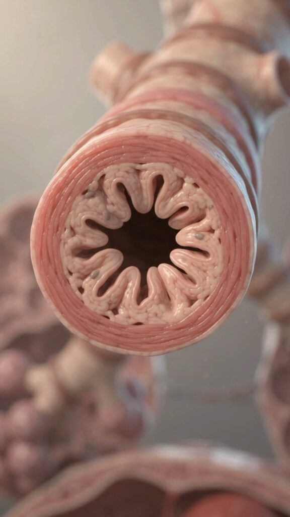
Why Asthma Makes Breathing Hard: Inside a Narrowing Airway
See how tight airway muscles, swelling and extra mucus can make breathing harder during asthma, and learn when symptoms need emergency care.
Read the story →
What Actually Happens Inside Your Brain During a Stroke?
One blocked artery can disrupt a brain network. Follow the chain from blood flow to neurons, then learn why possible stroke signs need emergency care.
Read the story →